By Yolande Lai Li-Ching and Yap Zhi Jiun (final year students, Malaysia) & Dr Shahir Ahmad (Neurology registrar, Malaysia)
By the time this gets published, I imagine Yolande and Zhi will be qualified. Sorry guys! My fault!!
Introduction
Movement disorders involve either too much or too little movement.
 Vs
Vs  Source: Google image
In more descriptive words, movement disorders are conditions that include involuntary improvement that are abnormal in initiation, implementation, velocity, frequency or posture.
One of the most common, and important, is Parkinson’s disease (PD), where there is a mixture of too little (bradykinesia) and too much movement (tremor and drug-induced dyskinesia).
The details of PD can be found here –> Parkinson’s disease
What we will be discussing instead are “hyperkinetic movement disorders”.
The common (essential) hyperkinetic movement disorder Vs less common (nice to know) are as follows.
| Common | Less common |
| Chorea
Dystonia Myoclonus Restless legs syndrome Tremor Tics
|
Abdominal dyskinesia
Akathitic movements Ataxia/asynergia/dysmetria Athetosis Ballism Hemifacial spasm Hyperekplexia Hypnogenic dyskinesia Jumping disorders Jumpy stumps Movement of toes and fingers Myokymia and synkinesis Myorhythmia Paroxysmal dyskinesia Periodic movements in sleep REM sleep behavior disorder Stereotypy |
Building the whole picture:
Do not panic when you come across any movement disorders cases in your future practice or in your coming exams.
The Four steps systematic approach will help you to get clearer overall picture, or at least gain good marks in your exams.
STEP 1: Always describe the movement that you observed. This is also known as
phenomenology.
What kind(s) of involuntary movement(s) is/are present?-What is the nature of any improvement of movement?
*Table 2 might help you a bit
(NB. Often this is straightforward but sometimes the movements do not fall neatly into a single descriptive category and combined terms are used eg, myoclonus-dystonia)
STEP 2: Are there any associated neurological symptoms/clinical syndromes?
What mix of phenomenology is present?-What other neurological or systemic features are present?
(NB. Abnormal movements can be part of a more widespread neurological disorder or a complication of a previously diagnosed condition eg. Tremor in multiple sclerosis)
STEP 3: Aetio-Pathological diagnosis Are there any other medical issues? eg hyperthyroidism, Wilson’s disease, renal or liver failure, metabolic problems or any relevant drugs currently or in the past (Eg. β-agonists, antipsychotic medication, antiemetic)
STEP 4: Could it be genetic?
Drawing a family tree if there is family history would be extremely helpful
Table 2: Characteristics of the Common Types of Hyperkinetic Disorders
| Chorea | Involuntary, irregular, purposeless, nonrhythmic, often abrupt, rapid, and unsustained movements seem to flow randomly from one body part to another; they are unpredictable in timing, direction, and distribution. |
| Dystonia | Both agonist and antagonist muscles of a body region contract simultaneously to produce a twisted posture of the limb, neck, or trunk. In contrast to chorea, dystonic movements repeatedly involve the same group of muscles. They do not necessarily flow or affect different muscle groups randomly. |
| Myoclonus | Sudden, brief, shock like jerks are caused by muscular contraction (positive myoclonus) or inhibition (negative myoclonus, such as asterixis). |
| Restless legs syndrome | An unpleasant, crawling sensation in the legs (or arms), particularly during sitting and relaxation, is most prominent in the evening (but can also occur during the day). It disappears (or is significantly relieved) during ambulation. |
| Tics | Abnormal, stereotypic, repetitive movements (motor tics) or abnormal sounds (phonic tics) can be suppressed temporarily but may need to be “released” at some point. The release provides internal “relief” to the patient until the next “urge” is felt. |
| Tremor | An oscillatory, usually rhythmic (to-and-fro) movement of one or more body parts, such as the neck, tongue, chin, or vocal cords or a limb. The rate, location, amplitude, and constancy vary depending on the specific type of tremor. |
Source: Fernandez, Hubert H, A Practical Approach to Movement Disorders, Diagnosis and Management, Second edition, Demos Medical Publishing; 2015:11
How really common it is?
Movement disorders are far more common than we thought. The real incidence worldwide may not reflect the true numbers as it may be under the radar. The most common movement disorders are restless legs syndrome and followed by essential tremor.
The table shown below is to give some rough idea on the overall prevalence worldwide . 
Source: Schrag A. Epidemiology of movement disorders. Parkinson’s Disease and Movement Disorders. 4th ed. Philadelphia, PA: Lippincott Williams & Wilkins; 2002:73–89
The spectrum of hyperkinetic movement disorders
Movement disorders present in a wide clinical spectrum rather than as a single distinctive entity.

N.B : *Tics are suppressible compared to other movement disorders Bear in mind, functional movement disorders can present with any form of this wide spectrum hyperkinetic movement disorders.
To make things sometimes far more complicated, sometimes a single description may not be enough as a few may coexist and its blend into one another. For example, Ballismus, Chorea, Athetosis and Dystonia often co-exist.

Now we shall discuss the common movement disorders and a few example of common related diseases.
1. Tremor
Now try to describe the phenomenology that you see in this video :
Tremor is the second most common involuntary movement disorder seen in clinical practice.
In the descriptive way, tremor is defined as oscillatory, usually rhythmic (to-and-fro) movement of one or more body parts, such as the neck, tongue, chin, or vocal cords or a limb.
The rate, location, amplitude, and constancy vary depending on the specific type of tremor.
Table 1: Different types of tremor and classification
| Types of tremor | Descriptions | Seen in |
| Resting tremor | occurs when the affected extremity is at complete rest and diminishes with movement | Parkinson’s disease
Advanced Essential tremor |
| Postural tremor | occurs when the affected limb is held in position against gravity. | Essential tremor
Medication induced tremor |
| Action or kinetic tremor | occurs during voluntary movement. | Essential tremor
|
| Intention tremor | marked increase in tremor amplitude during the terminal portion of a targeted movement | Cerebellar tremor |
| Task-specific tremor | emerges during a specific activity. | primary writing tremor |
Source: Fernandez, Hubert H et al, A Practical Approach to Movement Disorders, Diagnosis and Management, Second edition, Demos Medical Publishing (2014)
The next diagram provides a more complete guide to the approach of tremor.

Source: Bhatia KP, Bain P, Bajaj N, Elble RJ, Hallett M et al, on behalf of the Tremor Task Force of the International Parkinson and Movement Disorder Society. Consensus statement on the classification of tremors. Mov Disord 2017, in press

Source: Bhatia KP, Bain P, Bajaj N, Elble RJ, Hallett M et al, on behalf of the Tremor Task Force of the International Parkinson and Movement Disorder Society. Consensus statement on the classification of tremors. Mov Disord 2017, in press
In summary……..

Essential tremor (ET)
It is the most common type of tremor. It is progressive, often inherited and usually begins in later adulthood.
ET is a slowly progressive 5-8 Hz tremor that is postural and worse on action, involving the hands, arms and may also affect the head, jaw, voice and tongue.
The tremor, usually symmetrical, is most noticeable when the arms are held up and during purposeful actions like eating, drinking from a cup, writing or finger nose testing.
There may be a yes–yes head tremor, although head tremor alone is rare.
Interestingly, ET can respond well to alcohol (not to encourage alcoholism but to give some diagnostic value) and relaxation.
Diagnosis is an exclusion of alternatives:
- Normal tone
- Normal coordination
- No bradykinesia
- No exposure to tremorgenic medications*
- Normal investigations (thyroid function test, plasma caeruloplasmin and 24h urinary copper for Wilson’s disease in patients under 50 y/o)

Figure 1. Handwriting and spiral drawing by patient with essential tremor

Source : Elble RJ. Diagnostic criteria for essential tremor and differential diagnosis. Neurology 2000;54:S2–6

Source: R Bhidayasiri, Postgrad Med J 2005;81:756–762
Treatment
Treatment may not be needed for mild cases. This is mainly due to the potential side effects of the medications, especially when given for long term.
Different body parts a may also have different pharmacological responsiveness.
| Lifestyle modification | ● avoid caffeinated drinks
● non-medical relaxation techniques to reduce stress ● minimal amount of alcohol during social events (hmmm..) |
| Medication | ● 1st line:
○ Propanolol ○ Primidone ( old antiepileptic, rarely used nowadays) ● 2nd line ○ Gabapetin ○ Topiramate ○ Clonazepam ● 3rd line: ○ Clozapine |
| Surgery | 1.Deep brain stimulation of the ventral intermediate nucleus of the thalamus
2.Focused ultrasound device(latest therapy and technically and no knife/surgery involved |
The other common type of tremor is rest tremor which commonly seen in Parkinson’s disease. The details can be found on this page –> Parkinson’s disease
But for quick comparison, the table below help to highlight the differences between ET and resting tremor.

Adapted from: Hubert Fernandez, Andre Machado, Mayur Pandya et al, A Practical Approach to Movement Disorders, 2nd Edition, Diagnosis and Management- Demos Medical (2014)
Not to forget, the psychological tremor is another important and interesting tremor seen in the clinical practice.

Source: Manyam BV. Uncommon forms of tremor. In: Watts RL, Koller WC, eds. Movement disorders: neurologic principles and practice. 2nd ed. New York: McGrawHill, 2004:459–80
Other types of tremors that less common are:
| Other type of tremor | Presentation | Treatment |
| Dystonic
|
● Not rhythmic and tend to have a directional element, moving more in one direction.
● patients usually have a sensory trick to control the tremor (geste antagoniste) |
Botulinum toxin injection |
| Cerebellar | ● Slower (3 Hz) tremors that increases as the limb is moved towards the target (intention tremor).
● usually presented with clumsiness or unsteadiness, incoordination, gait ataxia and slurred speech (dysarthria) |
No medication found to be really effective |
| Orthostatic | ● a rare tremor that appears in the legs on standing. No difficulty in walking.
● Confirmation is by electromyography showing a 16 Hz pattern |
● Clonazepam
● Gabapentin ● Levodopa |
Other interesting but much rarer tremor includes:
Palatal tremor : https://www.youtube.com/watch?v=yps2i3mjbN4
Wing beating tremor (seen in late stage of Wilson’s disease) : https://youtu.be/JIFpgSt7awo
Holmes’ tremor : https://youtu.be/ithJUroqUs0
Dystonias
What is it?
Dystonia is a movement disorder characterized by sustained or intermittent muscle contractions causing abnormal, often repetitive, movements, postures, or both.
Dystonic movements are typically patterned, twisting, and may be tremulous.
Dystonia is often initiated or worsened by voluntary action and associated with overflow muscle activation pain in the contracting muscles and dystonic movements.
Anatomical distribution

Clinical features of isolated dystonias
|
The present symptoms: |
The absent symptoms: |
| •Sustained involuntary movement, sometimes overlying spasms
•Consistent directional quality •Involves same body region(s) •Enhanced or produced by activity of involved area •Varies with change in activity or posture •Sensory tricks (geste antagoniste) may reduce symptoms •May have associated tremor |
•Weakness
•Amyotrophy •Spasticity •Ataxia •Ocular abnormalities •Abnormal eye movements •Retinal changes •Cognitive impairment •Seizures |
Classification of Dystonia
The Dystonia Task force by the International Movement Disorder Society 2013 has simplified dystonia classification by using 2 axes:
Axis 1: Clinical Characteristic
Axis 2: Etiology

Source: Albernase et al, Phenomenology and Classification of Dystonia: A Consensus Update, Movement Disorders, Vol. 28, No. 7, 2013
Axis 2: Etiology
|
Inherited or Inherited | |||
| Inherited | Inherited | Idiopathic | ||
| Evidence of degeneration
|
Autosomal dominant
|
Perinatal brain injury | Sporadic
|
|
| Evidence of structural (often static) lesions | Autosomal recessive | Infection | Familial | |
| No evidence of degeneration or structural lesion | X-linked recessive
|
Drug | ||
| Mitochondrial | Toxic
|
|||
| Vascular | ||||
| Neoplastic | ||||
| Brain injury | ||||
| Psychogenic | ||||
Source: Albernase et al, Phenomenology and Classification of Dystonia: A Consensus Update, Movement Disorders, Vol. 28, No. 7, 2013

With advancing genetic sequencing, many genes have been identified that associate with certain types of dystonia. (but yet many gene are yet to be discovered) The list is quite long and exhausting, but you don’t have to know in details. The most important are Dopa-responsive dystonia (aka Segawa’s disease (DYT5a gene) which has a remarkable response with relatively small doses of levodopa and Oppenheim/Primary dystonia (DYT1 gene) which has potentially good response with deep brain stimulation therapy.
Dopa-responsive dystonia (Segawa disease) or DYT5.
Also known as hereditary progressive dystonia with diurnal fluctuations (Segawa et al, 1976). Age of onset is in childhood ( 1 to 12 years). Typically present with progressive dystonia manifesting as gait disturbance or foot dystonia and, less commonly, with craniocervical dystonia, including spasmodic dysphonia.
Initially, children show diurnal fluctuation, with progressive worsening of symptoms toward the end of the day, and are often remarkably normal in the morning.
If not recognized and treated, the diurnal characteristic may abate, and children develop features of Parkinsonism. Rarely, patients with DRD also may experience hypotonia and mild developmental delay.
The early onset of gait impairment leads to an erroneous diagnosis of cerebral palsy. Patients with DRD have a dramatic response to small doses of levodopa.
A trial of levodopa,one tablet (100 levodopa/25 carbidopa) for 5 to 7 days, is an effective and highly rewarding method of diagnosis. All patients remain levodopa responsive and levodopa dependent throughout their lives. Children typically need their dose to be adjusted as they grow older. However, adults derive benefit from the same dose for years, without any wearing off or dyskinesias.
Dystonia body distribution in quick glance and treatment
▶ Focal
| Type | Presentation | Treatment |
| Cervical dystonia [COMMON] | neck pain, torticollis (turning to one side), laterocollis (leaning to one side), anterocollis (forwards), retrocollis (backwards), tremor, hypertrophy of sternocleidomastoid or splenius capitis | botulinum toxin injection
anticholinergics |
| Blepharospasm [COMMON] | blinking and forced closure of both eyes, not painful | botulinum toxin injection |
| Spasmodic dysphonia [RARE] | strangulated speech, high pitched strangled quality | botulinum toxin injection into overactive laryngeal muscles |
| Oromandibular dystonia | mouth and tongue affected, jaw may close on protruded tongue | botulinum toxin injection |
| Task specific dystonia
eg. writer’s cramp |
Difficulty in performing specific complicated motor task, often related to patient’s occupation | Change in technique of carrying out task
Botulinum toxin injection only gives limited improvement |
▶ Segmental; for example, Meige’s syndrome— blepharospasm, with lower face and cervical dystonia or brachial dystonia. video: https://youtu.be/RfnLtt716vQ
▶ multifocal – Two noncontiguous or more (contiguous or not) body regions are involved.
▶ generalised – The trunk and at least 2 other sites are involved.
▶ hemidystonia—More body regions restricted to one body side are involved. Example: acquired brain lesions in the contralateral hemisphere
The classifications of dystonia are really important as it determines the treatment of choice.
For focal and segmental dystonias the treatment of choices involves botulinum neurotoxin injection, in which the side effect profile is far less common (safer) and tend to be more localized.
On the other hand, the treatment or generalized dystonias often involves medications or surgery (deep brain stimulation).
Key Investigations
- Details medical and family history
- Excludes Wilson disease (as it is potential treatable disease)
- 24 hr urinary copper
- Plasma caeruloplasmin
- Slit lamp examination for Kayser-Fleisher rings
- Genetic testing
- DYT1 (for young onset)
- Huntington’s disease and SCAs
- Full blood count and fresh blood film for acanthocytes
- MRI brain scan
- Therapeutic trial of levodopa
Myoclonus
Often happens in a boring lecture and then a sudden thunderbolt-like-jolt woke you up. You pretended nothing had happen but the lecturer obviously noticed that.. It felt as if you were “falling into the void”.
All of us experienced the hypnic jerk at some point of time in our life, especially when we were on the edge of sleep and these are normal. It can occur without other neurological deficits (essential myoclonus).
By definition, myoclonus is is defined as a brief, shock like, jerky involuntary movement involving one muscle or a group of muscles.
This is how it look like : https://youtu.be/N01NidRGe24
Myoclonus is seen in several epileptic neurodegenerative conditions, including mitochondrial disorders (myoclonic epilepsy with ragged red fibers), Lafora body disease, neuronal ceroid lipofuscinosis, and in other lysosomal storage disorders.
When myoclonus is pathological it is more often a feature of an underlying disease than a primary disorder.
Myoclonus can be classified according to the distribution or origin of the myoclonus generator.
| Distribution | Origin of the myoclonus generator |
| Focal | cortical |
| Multifocal | subcortical |
| Segmental | brainstem |
| Generalized | Spinal |
| Peripheral |
Characteristics that differentiate anatomical subtypes of myoclonus

Zutt, R. et al. Nat. Rev. Neurol. 11, 687–697 (2015)
Negative myoclonus: https://youtu.be/mMsrq8GQ8Bk
Brainstem myoclonus: https://youtu.be/xCBpig4qVG4
Propiospinal myoclonus: https://youtu.be/Oha-0w0oBmk


Teesta Soman, Anthony E. Lang, Movement disorder,Continuum Lifelong Learning Neurol 2009;15(6):167–190.

Zutt, R. et al. Nat. Rev. Neurol. 11, 687–697 (2015)
Investigation is directed by the clinical situation, and management is usually of the underlying disorder.
Key investigations:
EEG
MRI of brain/spine
Urinary copper?
Treatment


Source: Fernandez, Hubert H et al, A Practical Approach to Movement Disorders, Diagnosis and Management, Second edition, Demos Medical Publishing (2014)
Myoclonus in epilepsy
Commonest: Juvenile myoclonic epilepsy
Rare: Progressive myoclonic epilepsies which patients present with myoclonus but later develop generalised seizures, ataxia and dementia.
Treatment: Sodium valproate is the first line medication. Alternatively, lamotrigine and levetiracetam are helpful
(NB. DO NOT prescribe carbamazepine, phenytoin or gabapentin as these medication will make the epilepsy worse)
Hyperekplexia
One form of interesting but rare (brain stem in origin ) myoclonus is Hyperekplexia.
Hyperekplexia is defined as a pathologic exaggeration of the normal startle response.
It is characterized by a reflex jerk to auditory, somatic, or visual stimuli that produces a sudden grimace; abduction of the arms; and flexion of neck, trunk, elbows, hips, and knees.
A classic feature is a prominent startle response to nose taps that does not suppress with repeated testing. In an older child or adult, exaggerated startle; hyperreflexia; abnormal, wide-based gait (probably compensatory because of repeated falls secondary to startle); tonic spasm; and paroxysms of jerking are present.
Hereditary hyperekplexia is caused by a mutation in the subunit of the glycine receptor, leading to a disruption in the chloride ion channel
Clonazepam can have a marked ameliorative effect on these symptoms.
Tonic laryngeal spasm may also occur, putting these patients at a risk of respiratory arrest. Seizures may also be present.

A novel eight-step diagnostic algorithm for myoclonus
Zutt, R. et al. Nat. Rev. Neurol. 11, 687–697 (2015)
Chorea……Ballismus……Athetosis
Interestingly, definition of Chorea has been unchanged since 19th century. Chorea derives from the Greek word choreia meaning dance.
Chorea is defined as an involuntary movement that is brief, irregular, nonrhythmic, non purposeful and flows from one body part to another in a random fashion.
Unlike dystonia which is predictable, chorea is unpredictable (important clue).
The movements typically last longer than myoclonus and are briefer than dystonia (although dystonia may be combined with chorea in some patients).
It is usually present at rest and may increase with distracting maneuvers such as counting or performing mental arithmetic. The movements may be partially suppressible or incorporated into voluntary movements to make them less conspicuous. They usually disappear in sleep.
Chorea can be unilateral or bilateral. It can affect all body parts: face, trunk, and limbs.
It may blend into:
Ballismus when the movements are of large amplitude and intensity, involving proximal muscle groups
Athetosis when slow writhing distal movement involved.

In adults, the most common form of chorea is drug induced chorea e.g Levodopa induced chorea in Parkinson’s patient – better known as dyskinesia.
Out of this context, the most common cause of acquired chorea is vascular disorder e.g stroke. Interestingly, in Asia, the most common cause of chorea in adult is endocrine causes e.g hyperglycaemia (particularly in type-II DM).
In term of genetic chorea, Huntington Diseases is the most important cause in any part of the world
It also can be classified to Primary and Secondary Chorea
In children, chorea is rare and autoimmune chorea responsible for most the cases.

Teesta Soman, Anthony E. Lang, Movement disorder,Continuum Lifelong Learning Neurol 2009;15(6):167–190.

Teesta Soman, Anthony E. Lang, Movement disorder,Continuum Lifelong Learning Neurol 2009;15(6):167–190.

Acanthocytes (spiculated red blood cell)
Source: Rola Zamel, Razi Khan, Rebecca L Pollex and Robert A Hegele – Abetalipoproteinemia : two case reports and literature review. Orphanet Journal of Rare Diseases 2008, 3:19.
How to treat Chorea?
The main goal of management is treatment of the underlying condition rather than of the chorea itself. This includes removal of the offending drug, normalization of glycemia, immunomodulatory therapy for autoimmune chorea, or removal of an underlying neoplasm.
Anti-choreic medication are
- Atypical neuroleptics (evidence is scared)
- Tetrabenazine(monoamine depleter) effective but costly and tolerability profile
- Pallidal deep brain stimulation (DBS)for pharmacoresistant chorea or ballismus with significant disability.
We will discuss about 3 types of chorea that are relatively common compared to other type of chorea.
1.Sydenham chorea aka St Vitus dance
This is the most common cause of childhood chorea and is autoimmune in origin.
Chorea usually begins within 2 to 6 months following group A β-hemolytic streptococcal pharyngitis. This is related to anti-dopamine receptor antibody.
The chorea is usually generalized, although 20% to 30% of patients have hemichorea. Distal musculature of the hands is affected with a ‘‘piano playing’’ pattern of repetitive hand movements. Motor impersistence is prominent, and the child is not able to maintain a firm grasp (milkmaid’s grip). Some patients are unable to maintain tongue protrusion (known as adder’s tongue or darting tongue). Emotional lability and behavioral changes, including obsessive compulsive behavior, often accompany this syndrome (basal ganglia dysfunction).
Although a monophasic illness in most children, approximately 30% of affected patients will experience recurrent symptoms even months or years after cessation of their initial illness. Female patients may have recurrence of symptoms many years later during pregnancy (chorea gravidarum) or when exposed to oral contraceptives or estrogen therapy.
Sydenham chorea is diagnosed by history and supported by the presence of an elevated antistreptolysin O titer, an elevated anti-DNAase B titer, and a positive streptozyme test or positive throat culture for streptococcus pyogenes.
The condition is usually self-limited and resolves within 6 months.
- Valproic acid or tetrabenazine may beuseful in severe or prolonged cases.
- Prednisone 2 mg/kg/d for 4 weeks wasfound to achieve a more rapid remission of symptoms (Paz et al, 2006).
- IV immunoglobulinhas also been administered (Garvey et al, 2005)in severe and protracted cases
Treatment must also be implemented to prevent rheumatic fever (oral penicillin for 10 days), and patients must be assessed for carditis. They should receive penicillin prophylaxis for dental or surgical interventions.
2. Benign hereditary chorea.
This is a nonprogressive chorea of childhood onset.
It is inherited as an autosomal dominant disorder, caused by a mutation in the TITF-1 gene on chromosome14.
The disease may start at any point from infancy to adolescence, is associated with progressive decline in function until the second decade, and then is usually static. Affected children may have delay in walking, are described as clumsy, and have frequent falls. The choreic jerks can affect any part of the body, including the face,tongue, neck, trunk, or limbs. The choreic movements vary greatly in frequency and severity even within the same family.
Some individuals have infrequent movements that may not be recognized by the affected person and are only identified by an experienced examiner. Others can be so severely affected thatthey are unable to do simple tasks such as holding a glass of water. The intensity and frequency of the movements tend to worsen with stress or excitement and cease during sleep. The movements have usually stabilized with no further progression, and in some families, improvement has occurred over time . MRI of the brain is normal. No treatment may be necessary in this condition.
The TITF-1 genetic mutation should also be considered in all children with chorea, mental retardation, hypothyroidism, and chronic lung disease.
3. Hungtington Disease

George Huntington in 1872 (Wikipedia.org)
Huntington disease (HD)is the most frequent cause of a hereditary neurodegenerative choreic syndrome. It is caused by a faulty gene on chromosome 4, producing a protein called Huntingtin which is thought to increase the rate of cell death in the brain.
It is relatively rare disorder and affects about 8,000 people in the UK. HD is an autosomal dominant neurodegenerative disorder (each child of an affected parent has a 50% chance of developing the disease)

The initial symptoms occur more frequently between the ages of 30 and 50, although onset can range from childhood/adolescence (juvenile form, also known as the Westphal variant) to individuals older than 70 years of age.
HD can present to the clinician with one of three symptom complexes classically described in this condition (motor/physical, cognitive, and neuropsychiatric/emotional) or in combination. The motor symptoms include chorea, dystonia, and tics.


Source: http://www.hda.org.uk
Parkinsonism usually develops later in the course of the disease.
The juvenile form of HD presents as a predominantly hypokinetic movement disorder with parkinsonism but also myoclonus.
How to confirm the diagnosis?
For HD, a trinucleotide cytosineadenosine-guanine (CAG) equal to or greater than 36 repeats is diagnostic in the presence of characteris tic symptoms.

Graphic showing the excessive repetitions of the cytosine-adenine-guanine (CAG) nucleotide sequence in a gene from a Huntington’s disease patient (bottom) compared to a gene from a person without the neurodegenerative disorder (top).
Credit: National Institute of General Medical Sciences, National Institutes of Health

A table summarizing the different possible results of a predictive HD gene test.
In the nutshell….

Source: Wikipedia.org & open.osmosis.org
The hallmark feature in clinical MRI found in adult-onset HD is caudate atrophy.

Teesta Soman, Anthony E. Lang, Movement disorder,Continuum Lifelong Learning Neurol 2009;15(6):167–190.
What can be offered to them?
No disease-modifying therapies are available for HD (although several trials are ongoing).
The current treatment are:
- Symptomatic/Supportive therapymainly to alleviate the symptoms
- Anti-choreic medication – atypical antipsychotic, tetrabenazine (tobe used with caution)
- Deep brain stimulation for pharmachological resistant chorea with significant disability
Tics and Tourrete’s syndrome

Georges Gilles de la Tourette (1857–1904), namesake of Tourette syndrome
Check out this video of a teenager with Tourette’s syndrome who sucesfully became a tv host!
A tic is an involuntary movement or vocalization that is usually of sudden onset, brief, repetitive, and stereotyped, but nonrhythmic.
Tics frequently imitate normal behavior, often occurring during normal activity and without alteration of consciousness.
Unlike most movement disorders, tics can persist during sleep.
A tic is usually associated with a premonitory “buildup” sensation or feeling of discomfort that is often localized to the affected area. Usually,the individual experiences a sensation of relief once the tic has occurred.
How to clarify tics?
Tics can be classified as motor, phonic or vocal.
Tics can also be categorized as simple or complex, depending on the manifestation

Source: Fernandez, Hubert H et al, A Practical Approach to Movement Disorders, Diagnosis and Management, Second edition, Demos Medical Publishing (2014)
There are few characteristics of tics that helps to differentiate from other movement disorders.
- Tics are commonly associated with a premonitory sensation
- Tics can be suppressed
- Typically, tics do not disrupt volitional movements.
- There can also be tic blocking, during which there is a sudden stoppage of movement.
- The severity of tics usually waxes and wanes
- Involvement in activities that require a great deal of attention or concentration usually diminishes the frequency of tics.
What is Tourrete’s syndrome? How does it different from tics?
GTS is found in all ethnic groups and occurs more frequently in males than females.
Tics and GTS may be more frequent in a special education classroom
The pathophysiology of GTS is thought to involve abnormalities in corticostriatothalamocortical circuitry.

Other behavioral abnormalities that have been associated with GTS include obsessive-compulsive disorder, attention deficit hyperactive disorder, separation anxiety, overanxious disorder, simple phobia, social phobia, agoraphobia, mania, major depression, and oppositional defiant behavior.
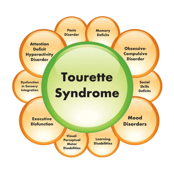
Tourette’s syndrome and other possible (under diagnosed) associated behaviors and psychiatric disorders
How to treat them?
The treatment of GTS is directed toward three goals:
- Control of motor and vocal tics,
- Management of behavioral disorders
- Treatment of psychiatric manifestations, ADHD, and OCD
In addition to pharmacologic interventions, behavioral treatments and psychiatric interventions may be very useful


Comella, Cynthia L. GILLES DE LA TOURETTE’S SYNDROME AND OTHER TIC DISORDERS, CONTINUUM: Lifelong Learning in Neurology: June 2004 – Volume 10 – Issue 3, Movement Disorders
In summary..

Teesta Soman, Anthony E. Lang, Movement disorder,Continuum Lifelong Learning Neurol 2009;15(6):167–190.
References
- Fuller G. Hyperkinetic movement disorders: Shakes, jumps and jolts. Practical Neurology. 2010;10(2):114-23.
- GILLES DE LA TOURETTE’S SYNDROME AND OTHER TIC DISORDERS. Comella, Cynthia L. CONTINUUM: Lifelong Learning in Neurology: June 2004 – Volume 10 – Issue 3, Movement Disorders
- Teesta Soman, Anthony E. Lang, Movement disorder,Continuum Lifelong Learning Neurol 2009;15(6):167–190.
- Schrag A. Epidemiology of movement disorders. In: Jankovic J, TolosaE, eds. Parkinson’s Disease and Movement Disorders. 4th ed. Philadelphia, PA: Lippincott Williams &Wilkins; 2002:73–89
- Fernandez, Hubert H, A Practical Approach to Movement Disorders, Diagnosis and Management, Second edition, Demos Medical Publishing; 2005:11
- Bhatia KP, Bain P, Bajaj N, Elble RJ, Hallett M et al, on behalf of the Tremor Task Force of the International Parkinson and Movement Disorder Society. Consensus statement on the classification of tremors. Mov Disord 2017, in press
- Elble RJ. Diagnostic criteria for essential tremor and differential diagnosis. Neurology 2000;54:S2–6.Zutt, R. et al. Nat. Rev. Neurol. 11, 687–697 (2015)
- Manyam BV. Uncommon forms of tremor. In: Watts RL, Koller WC, eds. Movement disorders: neurologic principles and practice. 2nd ed. New York: McGrawHill, 2004:459–80.
- www.hda.org.uk
- wikipedia.org